

What is the Retina?
The retina is a thin layer of nerve cells located at the back of the eye. The light that enters the front of the eye is focused in the retina and converted into electrical impulses. The optic nerve sends these impulses to the brain, where the visual information is processed and converted into images.
How the Retina System Works
There are several components that make the retina function. A disease or problem in any of these areas can cause vision loss.
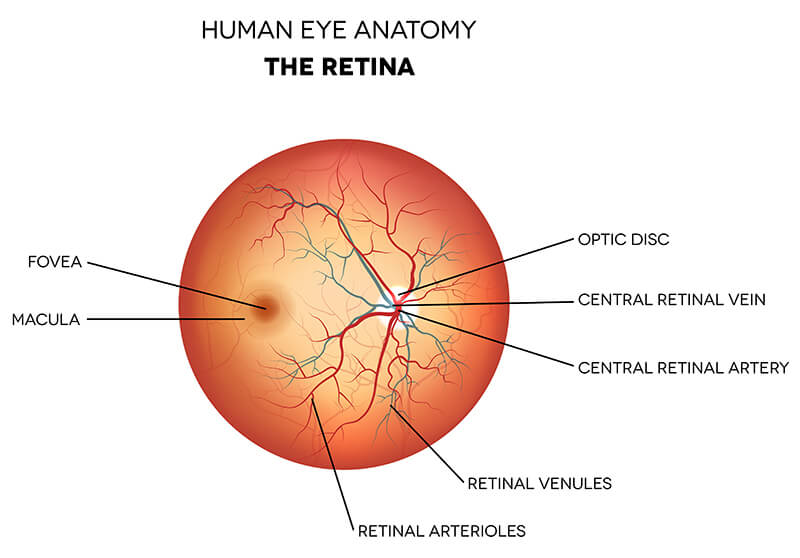
- Macula – a small area in the center of the retina that allows us to see detailed images clearly.
- Fovea – a small depression in the center part of the macula where visual acuity (sharpness) is highest.
- Peripheral Retina – the area outside the macula that enables side vision and night vision.
- Photoreceptors – cells in the eye that convert light into impulses that are sent to the brain. There are two types of photoreceptors: rods and cones. Rods give us night vision, and cones enable our color vision.
Retina Diseases and Conditions
There are many retina diseases and conditions, and some are very rare. Any patient who is at risk for retina problems, or who has already been diagnosed with one, should have regular dilated eye examinations and follow-up with an ophthalmologist in order to retain as much vision as possible.
Some of the more common conditions are described below:
Age-Related Macular Degeneration (AMD or ARMD)
This is a very common disease that causes central vision to deteriorate over time, which affects your ability to read, drive, or recognize faces. AMD affects the macula, the central part of the retina. The macula is responsible for your ability to see fine detail.
Central Retinal Vein Occlusion (CRVO)
This condition develops from a blood clot or blockage of the central vein that drains blood from the retina. It usually affects adults over 50 and occurs in one eye. CRVO often causes macular edema (swelling) and the growth of abnormal new blood vessels. The major symptom is blurred vision.
Central Serous Chorioretinopathy
This condition is caused by fluid leakage from a layer of tissue under the retina called the choroid. This results in distorted vision. It occurs most frequently in men ages 30-50 as a result of stress, hypertension, autoimmune disorders and use of steroids. Many cases resolve themselves within a month. If symptoms persist, photodynamic therapy or laser treatments are usually successful.
Diabetic Retinopathy
Diabetic Retinopathy affects the blood vessels in the retina of diabetic patients and can cause severe vision loss.
Epiretinal Membrane (ERM) also known as Macular Pucker
ERM happens when a defect in the retina causes semi-translucent membranes to form on the inside of the retina. In mild cases patients may not have symptoms, however, when the membranes affect the macula they can cause visual distortion in which straight shapes appear crooked. They often appear like cellophane, and cause tearing or puckering of the retina. There are no eyedrops or medications to treat ERM. In cases where vision loss is significant, vitrectomy surgery is the only recommended treatment.
Macular Edema
Macular edema is an extremely serious condition that can lead to severe vision loss and blindness. The edema is a blister of fluid that occurs in the layers of the macula. Leaking from damaged blood vessels or growth of abnormal new vessels causes the swelling. There are many underlying conditions that can create this, including diabetes, macular degeneration, inflammatory diseases such as uveitis, blood disorders, and eye injuries.
The most effective treatment for macular edema involves addressing the underlying cause of the macular edema whenever possible (an example would be controlling blood sugar levels in diabetes). Ophthalmologists use intravitreal injections as a direct method of controlling excess fluid caused by leaking blood vessels.
Macular Hole
With this syndrome, a hole or tear appears in the macula, and causes distorted or wavy vision. During the aging process the vitreous gel often begins to shrink and separate from the retina. If a piece of the vitreous sticks to the macula it can cause a hole to develop. This is a serious eye problem that can lead to blindness if not treated promptly.
A macular hole diagnosis requires vitrectomy surgery. The surgeon will remove the vitreous that is pulling on the macula, and then put a gas bubble into the eye. The bubble flattens the macula and holds it in place while the eye heals. The gas bubble dissolves within a few weeks.
Retinal Detachment
Retinal detachment occurs when the retina detaches from the back of the eye. The retina will not function properly when it is separated from its blood supply. Retinal detachments can occur suddenly for a variety of reasons. Symptoms include “floaters” in the eye, flashing light, and/or a shadow on the side of the eye that may progress towards the center of vision. Retinal detachments must be evaluated and treated as soon as possible to minimize severe vision loss.
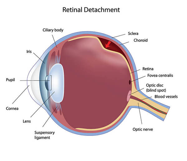
Laser treatments and pneumatic retinopexy can be used successfully for minor detachments. For more serious retinal detachments, vitrectomy or scleral buckle surgeries are necessary to repair the problem.